Investigations
There are several investigations important that are used in Ophthalmology to help diagnose and monitor eye conditions:
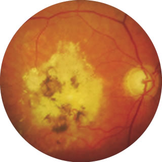
Retinal Photography
Optical Coherence Tomography (OCT)
Autofluorescence (AF) Imaging
Fluorescein Angiography (FFA) and Indocyanine Green (ICG) Angiography
Visual Field Testing
Retinal Photography
This involves using a special camera that allows accurate colour documentation of how the retina appears, which can be useful in documenting conditions and monitor any progress over time.
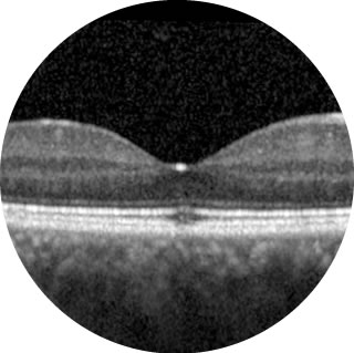
Optical Coherence Tomography (OCT)
OCT has transformed the understanding and treatment of retinal conditions in recent years. It is a non-invasive technique that captures specialised image of the retina, allowing individual retinal layers to be viewed. It is particularly useful in detecting fluid or bleeding that may occur in the retina in conditions such as Wet AMD, Diabetes and Retinal Vein Occlusion.
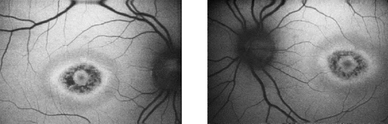
Autofluorescence (AF) Imaging
This is a non-invasive imaging technique that captures naturally emitted light wavelengths from the retina. It allows the detection of areas of retina that are damaged, and is useful in monitoring progression of retinal diseases.
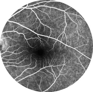
Fluorescein Angiography (FFA) and Indocyanine Green (ICG) Angiography
These are common diagnostics tests that involve the injection of a fluorescein (yellow) dye and/or indocyanine green dye into your bloodstream, through a vein in your arm. A series of photographs of the back of the eye are then taken, which can provide information on the blood supply affecting the retina, or the choroid – which is the blood supply beneath the retina. This is particularly useful in helping to diagnose leaking blood vessels in Wet AMD, and the extent of reduced blood supply in conditions such as diabetic retinopathy.
What are the risks of FFA or ICG Angiography?
- 1 in 10 patients may feel slightly nauseous, short of breath or develop a transient rash, but this rarely lasts for more than a few seconds.
- The fluorescein dye can give your skin a yellow tinge and make the colour of your urine yellow, but this usually settles after a day or two.
- There is an extremely rare (less than 1 in 200,000) risk of a severe allergic reaction to these dyes.
Visual Field Testing
This assesses the extent of your peripheral field of vision, which can be affected in conditions such as glaucoma. The test involves responding to small flashes of light which are targeted at various part of your peripheral vision and a map of your visual field is established.
Find out more…
To find out more about the eye, take a look at our patient leaflets or get in touch today. If you have any concerns about your eyes and require treatment, book an appointment with Venki Sundaram at one of our Hertfordshire clinics.